WHAT IS CELL ORGANELLE?
As we all know that ‘cell is the structural and functional unit of life’ and organelles are present within a cell. Cell organelles are present in every cell. The term organelle is derived from the word ‘organ’. Organelles mean “tiny organs”. Thus, we can describe cell organelles as tiny cellular structures present in the cytoplasm of both eukaryotic and prokaryotic cells. Few cell organelles provide shape and support, whereas, others are involved in the locomotion and reproduction of a cell. There are different organelles of cells, depending upon the presence/absence of membrane.
Different organelles of cell
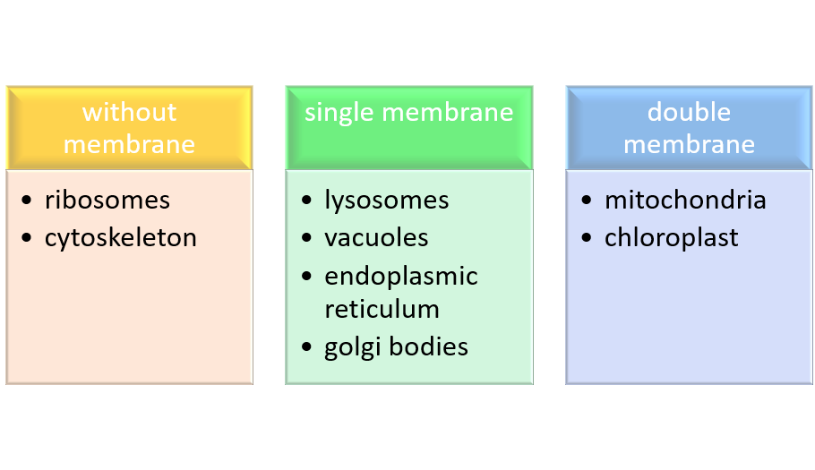
There are different organelles of cell and they can be with or without membrane.
- Cell wall
- Plasma membrane
- Nucleus
- Mitochondria
- Plastids
- Golgi apparatus
- Endoplasmic reticulum
- Ribosomes
- Lysosomes
- Cytoskeleton
- Vacuoles
- Peroxisomes
In this article, we will discuss only the structure and
Let’s dive in….
Cell wall
It is the outermost boundary in most of the prokaryotes and plant cells (eukaryote). The Prokaryotic cell wall is a semi-rigid, non-living component of the cell that surrounds the plasma membrane.
Structure
Prokaryotic cell wall:- Cell wall in prokaryotes comprises two heteropolymer, mucopeptide, and peptidoglycans (murein). Murein is a complex of disaccharides and polypeptide (NAM &NAG).
Both NAM (N-Acetyl Muramic Acid) and NAG (N-Acetyl Glucosamine) linked alternatively to form carbohydrate backbone like this;
-NAM-NAG-NAM-NAG-
A tetrapeptide chain is attached to NAM. In addition, adjacent tetrapeptide may be directly bound to each other or maybe linked by an oligopeptide bridge to form a cross-link between two chains.
Eukaryotic cell wall:- a typical plant cell has three distinct regions:
- Primary wall (thin and elastic)
- Secondary wall (thick and rigid)
- Middle lamella
The primary wall is present in growing cells (meristematic tissues in case of the plant cell), comprises 30-40% cellulose(dry weight), 50% of polysaccharides ( hemicellulose & pectin) and 5% glycoproteins rich in hydroxy proteins.
The secondary wall is generally very rigid and does not alter its shape. These are much thicker than the primary walls and consist of 40-45% cellulose, 15-35% hemicellulose, 15-30% lignin and negligible amounts of pectic polysaccharides. Moreover, the Secondary wall is deposited on the primary wall, towards the cytoplasm. It consists of cellulosic chains that bundle together to form micelles or elementary fibrils.
Middle lamella is present between the two adjacent cell walls. In other words, it is sandwiched between the primary and secondary walls. This middle lamella comprises pectin, cellulose, calcium, magnesium pectate, etc.,
The cell wall comprises polysaccharides that act as reserve carbohydrates for plants.
Plasma Membrane
It is a membrane present in all cells that separate the interior of the cell from the outside environment. In bacterial and plant cells, a cell wall is attached to the plasma membrane on its outside surface and the interior to the cell is the cytoplasm. Thus, the plasma membrane is present between the cell wall and cytoplasm of the cell in prokaryotes and plant cells. In the case of eukaryotes, the cell wall is absent. Therefore, the plasma membrane is the outermost boundary in such a case.
Structure
Among the various membranes of the cell, the plasma membrane typically has the clearest dark-light-dark or unit membrane appearance in electron micrographs. Several cell biologists attempted to understand the molecular structure of the cell membrane and proposed different model for the same. These models are classified into two basic categories:-
- Bilayer model
- Micellar model
In the
Mitochondria
‘Mitos’ means “thread” and ‘
Structure
Mitochondria has a sausage-shaped body having a thickness of about 0.5m. It is enveloped by two
The outer membrane shows a
The space enclosed by the inner membrane is called the matrix (inner chamber) and is usually filled up by a proteinaceous material. Within the matrix are small granules, small ribosomes, and a double-stranded circular DNA. The inner membrane is thrown into a series of irregular folds, called cristae or cristae mitochondrial. These cristae are tubular or finger-shaped and may be branched.
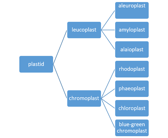
Plastids
These are double membrane-bound organelles present in most of the plant cells and some protozoans. They are generally absent in animal cells, bacteria, and fungi. Plastids
They are of two types:
- Leucoplast (colorless)
- Chromoplasts (color)
1. Leucoplast
They generally serve the purpose of storage. Generally, these are present in cells that do not receive direct sunlight. Leucoplasts are of three types:
- Aleuroplasts (store proteins)
- Amyloplast (store starch)
- Elaioplast ( store oil)
2. Chromoplast
They are colour plastids of various colour pigments. They act as a primary site for trapping and converting solar energy.
Based on their coloured pigment, chromoplast are of the following types:-
- Chloroplast (green)
- Phaeoplast ( brown)
- Rhodoplast (red)
- Blue-green chromoplast ( blue green)
Structure of Plastids (chloroplast)
Chloroplasts show great variation in shape, size, and numbers. Either they may have oval, disc-shaped or spherical structure or it may be band-shaped, ribbon-shaped.
Chloroplasts have a double-membrane envelope, 6-8nm thick each (phospholipid layer), consists of the inner and outer membrane. Further, the spaces between the double membranes covered with an aqueous matrix known as stroma having a thickness of about 10-20nm. This aqueous matrix contains various enzymes and proteins that are essential for cellular processes. The chloroplast shows a complicated lamellar system composed of small cylindrical structures called
In 1947, S. Granick and K. porter isolated chloroplast and showed that
The stroma of “matrix” of plastids usually contain several kinds of discrete bodies:-
- Starch grain (2mm in length)
- Postoglobuli (0.1mm in diameter) –a common feature of the chloroplast. Postoglobuli may accumulate colored pigment.
- Ribosomes (17 nm in diameter) –a
universal feature of thestroma . Fibrili (2.5 nm in diameter) – represent chromoplasts DNA.
Endoplasmic Reticulum
The Endoplasmic reticulum consists of a system of interconnected tubules with the membrane surrounding an interior cisternal space.
The Endoplasmic reticulum membrane is generally thinner than the
Typically, the cisternal space is 30nm in diameter but can increase considerably when induced by drugs. Due to its huge surface area, it is the largest membrane in the cell.
The outstanding morphological difference is the presence/ absence of ribosomes that are present/absent on their outer surface. Depending upon the presence/absence of ribosomes, it is of two types:-
- Rough ER (rough endoplasmic reticulum)
- smooth ER (smooth endoplasmic reticulum)
1. Rough ER
It is studded with numerous 18-22
2. Smooth ER
They lack ribosomes on their outer membrane. They appear to be more vascular with swollen cisternae. It occurs along with RER in some cell types, such as liver hepatocytes, but is more commonly proliferated in specialized cells lacking RER.
If you wanted to know about its role in protein synthesis and segregation, various protein modifications, then go ahead and Read more
Golgi Complex (editor of synthesized protein)
This is a membranous organelle present in the cell’s cytoplasm. Thus, the Golgi complex is an organelle that sorts and modifies proteins & lipids (fats).
Structure of the Golgi complex
Golgi apparatus appears as a complex array of interconnected cisternae, tubules & vesicles. Golgi apparatus made up of several fattened, stacked sacs referred to as cisternae. In addition, each Golgi body consists of 4-8 cisternae, slightly curved, each having a diameter of 0.5-1.0mm. The membrane of the cisternae is porous. Moreover, the pores may be localized or present all over the surface. Cisternae of the Golgi apparatus has two faces– cis and trans face.
The tubules arise from the peripheral region of the cisternae. The vesicles are goblet-like structures attached to the tubules.
The cis face convex in appearance is closer to the endoplasmic reticulum and acts as receiving compartment from the endoplasmic reticulum.
On the opposite side is the trans face, which is present towards the plasma membrane. In between cis and trans cisternae, the stack of cisternae known as medial cisternae.
For role in protein secretion, see: Golgi
Lysosomes
‘Lysis’ means digestion and ‘soma’ means body. Thus, lysosomes mean digestive bodies The lysosomes are the organelles that act as ‘digestive system’ of the cell. The lysosome is part of cells endomembrane system which also contains the ER, Golgi complex and associated coated and uncoated vesicles.
Lysosomes are most numerous in cells that take up macromolecules or larger substances from their environment. e.g., intestinal epithelial cells have several hundred lysosomes. These are absent in prokaryotic cells.
Lysosomes have their own distinguishing features. It is the ultimate destination of soluble proteins. Enzymes present in the
Structure of lysosomes
Lysosomes are dense, spherical vesicles and range in diameter from 0.2-0.8 mm. They have a single membrane, 10 nm in thickness, and comprises lipoprotein.
Although the lysosomal membrane is very delicate, it prevents the hydrolytic enzymes from reacting with the cytoplasmic substrates. Moreover, the membrane could disrupt by the blender or could breakdown on refrigeration.
The internal interior is quite heterogeneous ranging from a
There are two types of lysosomes:
- Primary
- Secondary
Primary lysosomes are pinched off from the trans cisternae of Golgi complex. Matrix is homogeneous and dense in primary lysosomes.
Endocytotic vesicles may fuse with the primary lysosome to form secondary lysosome. Their matrix is more heterogeneous.
Vacuoles and microbodies are also cellular organelles present inside a cell.
Also, see: Lysosomes, Vacuoles, and Microbodies
Nucleus
It is the most prominent cell organelle and was first discovered by Robert Brown in 1831. The nucleus contains the blueprint for determining the structure and function of cells. It is the largest organelle present in the cytoplasm, being about 10/µm in diameter in non-dividing cells.
The Nucleus is present in all eukaryotic cells with some exceptions, e.g., mature erythrocytes (RBCs) in mammals, dividing cells (for a brief period of time), phloem cells, etc. It is also absent in prokaryotes (bacteria and blue-green algae)
The Nucleus is usually present at the centre of the cell. In some cases, it is peripheral or it may occupy various positions. Depending upon the type of cells, their number, size, and shape vary.
Structure
A typical nucleus shows the following components:
- Nuclear envelope
- Nuclear sap
- Chromatin
- Nucleolus
1.Nuclear envelope
This double membrane structure separated by the inter-membrane space of 10-50nm. The inner & outer membrane of the nuclear envelope has a thickness of about 70-80Å and is mainly comprises proteins & lipids.
The outer membrane is usually studded with ribosomes and often appear continuous with the membrane of rough ER. Further, the inner membrane is lined by a dense, fibrillar network, known as nuclear lamina of varying thickness. Chiefly, this nuclear lamina provides structural support to the nuclear envelope.
Double membranes fuse with each other at irregular intervals leading to develop nuclear pores. Each nuclear pore has a diameter of about 60-90nm. In addition, the nuclear pore contains a complex, basket-like apparatus called the nuclear core complex. This complex fills the
2. Nuclear sap
Nuclear sap is a semi-fluid, gel-like substance present in the nucleus. It is also known as nuclear matrix, karyoplasm or nucleoplasm (just like the cytoplasm found inside a cell, the nucleus contains nucleoplasm). It contains nucleoproteins, nucleic acid, enzymes, and minerals.
3. Chromatin
Chromatin is a complex of macromolecules
The key protein in chromatin is the histones (
Histones help DNA to organize into ‘bead-like structures’ called nucleosomes. The amount of DNA tightly folded with each nucleosome bead is approx. 200bp. However, the no. of bp associated with each bead varies from about 150-250.
A nucleosome is wrapped around a set of 8 histones called an octamer. The nucleosome can be further folded to produce chromatin fibre. Also, chromatin fibres are coiled and condensed to form chromosomes.
4. Nucleolus
It appears as a spheroid body within the nucleus of eukaryotic cells. Its size can vary from 1-5mm approx. the number of nucleoli per nucleus is quite variable.
There are two major components of the nucleus:-
- Dense granular component of about 150Å in diameter, which may be either scattered or localized in discrete regions around the periphery.
- A fibrillary component made up of fibrils having a length of about 50-80 Å. Nucleolus shows plasticity in structural organizations since it undergoes alteration under environmental stress.
Ribosomes
Ribosomes are ribonucleoprotein particles found in a variety of cells either in a
Structure
Each ribosome is composed of two non-identical subunits. Bacterial ribosomes are composed of two unequal subunits with a segmentation constant of 50S (large subunit) and 30S (small subunit) and a combined sedimentation constant of 70S. On the other hand, eukaryotic ribosomes are 80S particles with two subunits, the large is 60S and the smaller is 40S. Both subunit contain ribosomal protein rRNA.
A 3-D model of E.coli ribosome was proposed that shows a large subunit having three protuberances sticking out of its upper side, a large flattened area on one of its surfaces on which the smaller subunit resides.
The small subunit has a head and a body separated by a constriction. Further, the body occupies about two-third of the volume of this subunit.
Cytoskeleton
we have the skeleton system for support. Similarly, the cell has a cytoskeleton. There are three major types of structures in cytoskeleton:-
- Microtubules
- Microfilaments
- Intermediate filaments
1. Microfilaments
Microfilaments are the narrowest among these three types. It is 1-2 µm in length and 5-7 in diameter. Microfilaments of higher eukaryotes are made up of many linked monomers of a protein called actin. Because they are made up of actin monomers, they are also known as actin filament. Actin filament forms a double helix, 6 nm wide, with globular subunits 5.5 nm long and a 37 nm helix repeat. Like microtubules, microfilaments also have polarity.
2. Intermediate filament
These are smooth surfaced, solid and unbranched filaments having a diameter 7-11 nm that lies between those of microtubules and microfilaments. Unlike microfilament and microtubules, intermediate filaments are chemically heterogeneous in structure, composition, and solubility.
The basic unit of intermediate filament is the ‘two-chain coil’ which are identical ( homopolymer) and are formed from helix interactions. On the other hand, cytokeratin is heterodimers, with one chain of one type and the other of a different type. Once the dimer has formed, it interacts with a second dimer to form a tetramer(in parallel fashion), two of these units then associate in anti-parallel fashion to form a protofilament. Now, four protofilaments make up the 10 nm diameter filament seen in cells.
3. Microtubules
In 1800, microscopists have reported that cells and their extensions often contain a fibrous component. This was especially noticeable in cilia and flagella. In 1963, D. Slautterback coined the term microtubules. These are found virtually in all eukaryotic cells.
Structure
In animal cells, they usually radiate from a centrosome located near the nucleus while in plant cells they are more near the plasma membrane. They appear in longitudinal sections as long, thin rods or in cross-sections as hollow tubes ringed by 13 subunits. The outer and inner diameter of these tubules is 30nm and 14nm respectively. Therefore, wall thickness is 18nm.
A clear, unstained zone, 5-20nm in width, often surrounds the outer wall of the microtubule. The length of microtubules
They comprise polymers of tubulin protein subunits, which are of two types:
- α tubulin molecules
- β tubulin molecules
Also, see: Cytoskeleton
Vacuoles
Together with the presence of plastids and a cell wall, the vacuole is one of the three characteristics that distinguish plant cells from animal cells. They have a single membrane. Within many cells, up to 90% of the cell volume may be occupied by a single vacuole or multiple vacuoles. In meristematic tissue, several smaller vacuoles (
Vacuoles contain solutes in higher concentrations (0.4-0.6 M). For example, citrus fruits store approximately 0.3 M citric acid in vacuoles which can be squeezed out as juice. This compartmentation, therefore, prevents many important enzymes from being denatured.
Peroxisomes
They are single membrane-bound organelles that participate in oxidative reactions. Originally, Peroxisomes are organelles that carry out oxidation reactions leading to the production of hydrogen peroxide. Because H2O2 is harmful to the cell, peroxisomes also contain the enzyme catalase, which decomposes H2O2 either by converting it to H2O or by using it to oxidize another organic compound.
They are particularly abundant in plants cells. Peroxisomes do not have their own genomes and, therefore, synthesize their proteins (peroxins) from the nuclear genome. Most peroxins are synthesized on free ribosomes and then imported into peroxisomes as completed polypeptide chains.
Functions of different cell organelles
Specific organelle performs specific functions inside the cell. Let’s discuss the function of each organelle below:
1. The functions of a cell wall
- The Cell wall is rigid which means ‘it provides shape to the plant cell’.
- It acts as a physical barrier protecting cells from invading viruses, fungi, bacteria, etc. Thus, we can say that they serve as a part of a defence mechanism.
- It is semipermeable hence, allows the circulation of materials such as water, molecular nutrients, and minerals.
- It presents a barrier to cell expansion during cell division.
2. Functions of the Plasma Membrane
- It provides a framework in which components can be organized.
- It acts as a selectively permeable membrane. The Plasma membrane allows the cell to exchange material and information.
- It also acts as a site where energy is transduced (convert) from one type to another.
- Membranes also provide mechanical strength and electrical insulation.
3. Functions of Mitochondria
- The mitochondria transduce chemical energy used by the cell in synthetic reaction and active transport.
- Mitochondria store and transport ATP molecules.
- Mitochondria also involved in the biogenesis of haem.
- The proton gradient set up by electron transport can produce heat in newborns, hibernating animals and cold stressed animals.
4. Functions of chloroplasts (plastids)
- Photosynthesis: major function of the chloroplast is to convert light energy into chemical energy.
- Plastids serve the purpose of the manufacturing and storage of protein, starch, and oil.
- Plastids provide colour to fruits and flowers.
5. Functions of Endoplasmic Reticulum
- It plays a major role in the synthesis of the secretory proteins and their modifications.
- ER plays an indispensable role in the synthesis of glycogen, lipids, cholesterol and steroid hormones.
- It helps in the formation of a nuclear envelope and other cell organelles.
- It plays a major role in detoxification of certain drugs and muscle contractions.
6. Functions of Golgi Complex
- Golgi complex is the site of synthesis and concentration of glycoproteins and mucopolysaccharides.
- In plant cells, it plays an indispensable role in the formation of cell plate during mitosis.
- It associates with the formation of melanin
- Golgi complex is responsible for the
modification of non-secreted proteins.
7. Functions of Lysosomes
Lysosome performs two major functions:
- Phagocytosis: intracellular digestion of extracellular macromolecules (food or any other foreign substance)
- Autophagy: (self-feeding) intracellular degradation of organelles or macromolecules. At the times of starvation, some of the cell organelles like mitochondria, ER, etc. can broken down and digested by lysosomal enzymes to release energy without damaging or killing the cells.
8. Functions of Vacuoles
- Vacuole contains a variety of hydrolytic enzymes. In addition to hydrolases, plant vacuoles may contain inhibitors of serine endopeptidases.
- The second major function of plant vacuole is solute accumulation. These solutes include ions (Cl-, K+, and Na+ ), amino acids, sugars, organic acids (malate and citrate), and secondary metabolites. Vacuole synthesizes Some of these molecules, but most are import from the cytoplasm.
- The internal acidic environment of a vacuole aids in the degradation of larger molecules sent to the vacuole for destruction.
- Vacuoles remove potentially toxic substances from the cytosol, such as excess heavy metals and herbicides.
- They also aid in the lysis and recycling of misfolded proteins that have begun to build up within the cell.
9. Functions of Microbodies
These are of two types:
- Peroxisomes
- Glyoxysomes
Functions of Peroxisomes
The peroxisomes play an important role in photorespiration ( light-stimulated production of CO2). Photorespiration is a wasteful process for the plant cell, since, it significantly reduces the efficiency of the process of photosynthesis (i.e., it returns a portion of fixed CO2 to the atmosphere).
Functions of glyoxysomes
- One of the main functions of glyoxysome is to convert stored lipids into hexose sugars. This conversion of fat to sugar is known as gluconeogenesis. This hexose sugar is utilized by the emerging seedling which uses it as an energy and carbon source until it is able to produce its own sugar by photosynthesis.
- Glyoxysomes also function in nitrogen fixation and photorespiration.
- In the glyoxysomes, the glyoxylate cycle converts acetyl-coA to succinate for the synthesis of carbohydrates.
10. Functions of nucleus
- The nucleus has immense importance in the storage and utilization of genetic information. It is the repository of most of the cell’s genetic information, passing it on to the daughter cells in the course of cell division. The total genetic information stored in the nucleus is referred to as the nuclear genome.
- Nuclear envelope of nucleus regulates the exchange of electron, ions and molecules of various compounds.
- It controls the ongoing activities of the cell by determining which RNA and protein molecules are produced by the cell and when they are produced.
11. Function of the Ribosomes
Ribosomes carry out two main functions inside a cell
- They decode the genetic code into amino acid sequences.
- They form the peptide bonds to build protein.
12. Functions of Cytoskeleton
The cytoskeleton is of three major types:
- Microfilament
- Microtubules, and
- Microbodies
Functions of microfilament
- Actin filaments are involved in localized cell membrane movement.
- Microfilament also mediates the cytoplasm streaming or cyclosis in most of the cells.
- Another example of actin is the involvement of the sperm of an animal. When the sperm encounters the outer jelly coat of the egg, Ca2+ enters the sperm, causing the acrosome membrane to fuse with the plasma membrane. As a result, the coat is dissolved and the plasma membrane of sperm and the egg can fuse to form a zygote.
Functions of Intermediate filaments
Intermediate filaments play a significant role as motility molecules within the cytoplasm of different classes of cells.
- The keratin filaments of epithelial cells form a network around the nucleus and also ramify through the cytoplasm where their role is to hold the organelles into place.
- Desmin ( in skeletal muscles) filament forms an organized network at the Z-line where they help to integrate muscle function.
- Neurofilaments play an integral part in axon transport of different macromolecules including synaptic protein towards the presynaptic membrane.
- Intermediate filaments provide the clues to the clinicians with cell markers to determine the identity of the tumor.
Functions of Microtubules
- microtubules provide an internal skeleton or scaffold that provides structural support and helps to maintain the position of cytoplasmic organelles.
- They serve as the primary components of the machinery responsible for mitosis and meiosis.
- Microtubules form principal elements of cilia and flagella as motile cell projections.
REFERENCES
Cell biology: organelle structure and function by David E. Sadava. Page no.241-263, page no. 295-303, chapter 9
Cell biology by David E. Sadava; CBS publisher; from page no. 213-225
Cell and molecular biology by Prakash S. Lohar; MJP Publishers; from page no. 16-18, 96-98, 115-117, 122-130
The cell: A molecular approach by Geoffrey M. cooper, fourth edition
Cell biology, genetics, molecular biology, evolution and ecology by P.S Verma and V.K. Aggarwal; page no. 146-148, 186-190
Essau’s Plant Anatomy, third edition, chapter: The Protoplast: Plasma Membrane, Nucleus, and Cytoplasmic Organelles
Plant Biochemistry edited by P.M. Dey and J.B. Harborne, chapter no. 1: The plant, the cell and its molecular component
Essentials of biology, fifth edition by Sylvia S. Mader and Michael Windelspecht
Lehninger; principles of biochemistry by Michael M. Cox and David E. nelson, page no. 246,247, 795-96.
External links
https://academic.oup.com/jb/article-abstract/137/2/115/873179?redirectedFrom=fulltext