DNA Repair Definition
DNA repair is a collection of cellular responses by which a cell identifies and corrects any damage to the DNA molecules that encode its genome. Radiation, chemical mutagens, heat, enzymatic errors, and spontaneous decay constantly damage DNA.
Fortunately, much of the damage done to DNA by spontaneous chemical reactions in the nucleus, chemical mutagens, and radiation can be repaired.
DNA Repair Mechanism
Repair mechanisms that include nucleotide removal utilize a common four-step pathway:
- Detection: The damaged section of the DNA is recognized.
- Excision: DNA-repair endonucleases nick the phosphodiester backbone on one or both sides of the DNA damage and remove one or more nucleotides.
- Polymerization: DNA polymerase adds nucleotides to the newly exposed 3′-OH group by using the other strand as a template and replacing damaged (and frequently some undamaged) nucleotides.
- Ligation: DNA ligase seals the nicks in the sugar-phosphate backbone.
Generally, DNA Repair systems have five broad categories:
- damage reversal (light-dependent repair or photoreactivation),
- excision repair,
- double-strand break repair,
- post replicative repair, and
- the error-prone repair system (SOS response)
E. coli cells possess five well-characterized DNA repair mechanism. Mammals seem to possess all of the repair mechanisms found in E. coli except photoreactivation. Because most mammalian cells do not have access to light, photoreactivation would be of relatively little value to them.
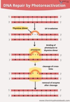
1.Light-dependent repair or Photo-reactivation
Exposure of DNA to Ultraviolet (UV) light causes linkage, or dimerization, of adjacent pyrimidines (cytosine and thymine) in DNA. These can be repair in several different ways. The simplest is to reverse the dimerization process and restore the original unlinked thymines. Light-dependent repair or photoreactivation of DNA in bacteria is carried out by a light-activated enzyme called DNA photolyase. DNA photolyase recognizes and binds to thymine dimers in DNA, and uses light energy to cleave the covalent cross-links.
Photolyase can bind to thymine dimers in DNA in the dark, but it cannot catalyze the cleavage of the bonds without light energy. Photolyase also splits cytosine dimers and cytosine-thymine dimers. The enzyme then falls free of the DNA. This enzyme thus reverses the UV-induced dimerization.

2. Excision Repair
Excision repair refers to the general mechanism of DNA repair that works by removing the damaged portion of a DNA molecule.
Steps
DNA repair mechanism of Excision repair involves at least three steps:
- A DNA repair endonuclease or endonuclease-containing enzyme complex recognizes and binds to the damaged bases in DNA. After that, it excises the damaged bases in DNA. As a result, gaps will form in the DNA strand.
- A DNA polymerase fills in the gap by using the undamaged complementary strand of DNA as template.
- Lastly, DNA ligase seals the break left by DNA polymerase to complete the repair process.
Excision repair are of two major types:
- Base excision repair
- Nucleotide excision repair
2.1 Base Excision Repair
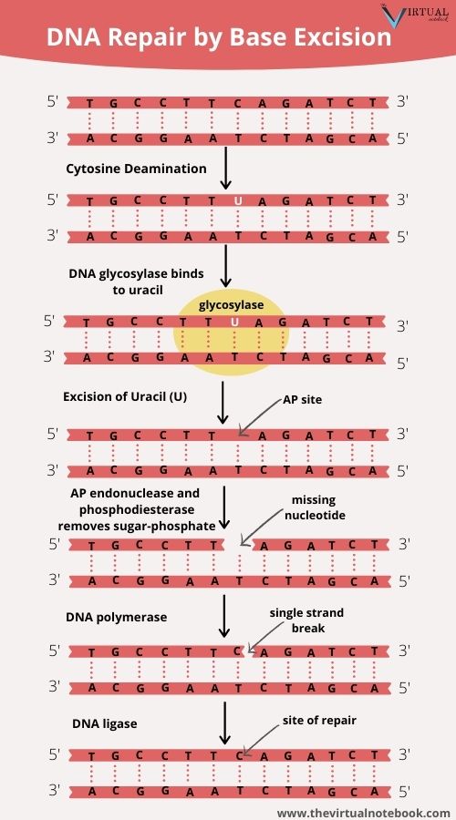
Base excision DNA repair pathways remove abnormal or chemically modified bases from DNA. It occurs in any of several ways:
- either by direct action of an agent such as radiation,
- or by spontaneous hydrolysis,
- by an attack of oxygen free radicals, or
- by DNA glycosylases,
In base-excision repair, a modified base is first excised and then the entire nucleotide is replaced. The excision of modified bases is catalyzed by a set of enzymes called DNA glycosylases, each of which recognizes and removes a specific type of modified base by cleaving the bond that links that base to the 1′-carbon atom of deoxyribose sugar.
Currently, at least five DNA glycosylases are known. Each glycosylase recognizes a specific type of altered base, such as deaminated bases, oxidized bases, and so on.
The glycosylases cleave the glycosidic bond between the abnormal base and 2-deoxyribose, creating apurinic or apyrimidinic sites (AP sites) with missing bases. An AP endonuclease then senses the minor distortion of the DNA double helix and initiates excision of the single AP nucleotide. DNA polymerase then replaces the missing nucleotide according to the specifications of the complementary strand, and DNA ligase seals the nick.
2.2 Nucleotide Excision Repair
Nucleotide excision DNA repair pathways remove larger defects like thymine dimers and bases with bulky side-groups from DNA. Nucleotide-excision repair is quite versatile and can repair many different types of DNA damage. It is present in cells of all organisms and is among the most important of all repair mechanisms.
In this DNA repair, a unique excision nuclease activity produces cuts on either side of the damaged nucleotide(s) and excises an oligonucleotide containing the damaged base(s). This nuclease is called an excinuclease (other than endonucleases and exonucleases). After that, helicase enzymes peel the part of the damaged strand and DNA polymerase fills the gap. Lastly, DNA ligase seals the DNA strand.
Like base excision repair, nucleotide excision repair is present in all organisms. Yeast involves approximately twelve genes whereas human beings involve twenty-five proteins.
3. Double-Strand Break Repair
Some damage to DNA is capable of breaking both strands of the double helix. There are two major pathways for repairing double-strand breaks:
- Either it can simply bring the ends back together (nonhomologous end joining), or
- it can use a mechanism that relies on the nucleotide sequences of a homologous piece of DNA, such as a sister chromatid or a homologous chromosome.
3.1 Non-homologous end joining
Non-homologous end-joining is a pathway that repairs double-strand breaks in DNA. We call it “non-homologous” because the break ends are directly joined without the need for a homologous template. This pathway often occurs when the cell is in G1 and a sister chromatid is not available for repair through homologous recombination. Nonhomologous end joining uses proteins that recognize the broken ends of DNA, bind to the ends, and then joins them together. If more than two broken ends are present, incorrect attachments can take place. Nonhomologous end joining is more error-prone than homologous recombination and often leads to deletions, insertions, and translocations(Various types of mutations).
See also: Types of mutations
3.2 DNA repair by homologous recombination
DNA repair by homologous recombination occurs in the cells when there is a homologous piece of DNA present in the nucleus, mostly in G2 and S phase of the cell cycle. Homologous recombination repairs a broken DNA molecule by using the identical or nearly identical genetic information contained in another DNA molecule, usually a sister chromatid. DNA repair by homologous recombination begins with the removal of some nucleotides at the broken ends, followed by strand invasion, displacement, and replication.
Two enzymes that play a role in homologous recombination are BRCA1 and BRCA2. The genes for these proteins will frequently mutate in breast cancer cells.
4. Post Replicative Repair
Sometimes DNA damage persists rather than being reversed or removed, but its harmful effects may be minimized. This often requires replication across damaged areas, so the process is called post-replication repair. For example, when DNA polymerase reaches a damaged site (such as a pyrimidine dimer), it stops the synthesis of the strand. As a result, it leaves a gap in the DNA strand. If allowed to remain, this gap will result in deficient and broken DNA.
The gap can be filled by strand exchange with the parental strand that has the same polarity, and the secondary gap produced in that strand can be filled by repair synthesis. The products of this exchange and resynthesis are two intact single strands, each of which can then serve in the next round of replication as a template for the synthesis of an undamaged DNA molecule.
5. The Error-prone Repair System (SOS response)
SOS repair is a complex set of processes that includes a bypass system that allows DNA replication to take place across pyrimidine dimers or other DNA distortions but at the cost of the fidelity of replication. Even though intact DNA strands will form, the strands are often defective. Emphatically, SOS repair is an error-prone system for DNA repair. This error-prone repair system eliminates gaps in the newly synthesized strands opposite damaged nucleotides in the template strands but, in so doing, increases the frequency of replication errors.
This repair system works in E.coli and other related bacteria. A significant feature of the SOS repair system is that it is not always active but induced by DNA damage. Once activated, the SOS system allows the growing fork to advance across the damaged region, adding nucleotides that are often incorrect because the damaged template strand cannot be replicated properly.
The SOS response appears to be a somewhat desperate and risky attempt to escape the lethal effects of heavily damaged DNA. When the error-prone repair system is operative, mutation rates increase sharply.
Other DNA Repair Mechanism
During the last few years, research on DNA repair mechanisms has demonstrated the presence of an army of DNA repair enzymes that constantly scan DNA. New results of this work have shown that several previously unknown DNA polymerases play critical roles in various DNA repair processes. In this section, we introduce a number of other processes that are important in the repair of damaged DNA.
Mismatch Repair
DNA mismatch repair (MMR) is a system that recognizes and repair erroneous insertion, deletion, and misincorporation of bases that can arise during DNA replication and recombination, as well as repairing some forms of DNA damage. In addition, the mismatch-repair system corrects small unpaired loops in the DNA, such as those caused by strand slippage in replication.
Mismatch repair encompasses about 99% of all DNA repairs. Mismatches often involve the normal four bases in DNA. For example, a T may be mispaired with a G. Because both T and G are normal components of DNA, mismatch repair systems need some way to determine whether the T or the G is the correct base at a given site. The repair system makes this distinction by identifying the template strand, which contains the original nucleotide sequence, and the newly synthesized strand, which contains the misincorporated base.
Steps
- When a mismatch repair system detects a mismatch, it cut out a section of the newly synthesized strand in two places and removes a region around the mismatch. The excised region is variable in size.
- Then an exonuclease degrades the cleaved DNA strand to a position on the other side of the mismatch (the excision step), creating a single-stranded gap.
- After excision, DNA polymerase and DNA ligase fill in the gap by using the remaining strand as a template.
The proteins that carry out mismatch repair in E. coli differentiate between old and new strands by the presence of methyl groups on special sequences of the old strand.
SOURCES AND EXTERNAL LINKS
Principles of Genetics by Robert H. Tamarin, 7th edition; chapter 12: DNA: Its Mutation, Repair, and Recombination.
Genetics Essentials: concepts and connections by Benjamin A. Pierce, chapter 13: Gene Mutations, Transposable Elements, and DNA Repair.
Principles of Genetics, 6th edition by D. Peter Snustad and Michael J. Simmons, Chapter 13:Mutation, DNA Repair, and Recombination
Also, Genetics: A Conceptual Approach by Benjamin A. Pierce , fourth edition chapter 18: Gene Mutations and DNA repair
Lastly, Genetics Principles and analysis Fourth Edition, Daniel L. and Hartl Elizabeth W. Jones, Chapter 13:Mutation, DNA Repair, and Recombination
https://en.wikipedia.org/wiki/Homology_directed_repair
https://en.wikipedia.org/wiki/Non-homologous_end_joining
https://en.wikipedia.org/wiki/DNA_mismatch_repair
Hi
Hello Anil