WHAT IS MEIOSIS?
Meiosis is a specialized kind of cell cycle that reduces the chromosome number by half, resulting in the production of haploid daughter cells. In addition, Meiosis includes two sets of divisions that distribute the haploid set of chromosome and ultimately results in four cells. Moreover, the term meiosis was coined by J.E. More and J.B. Farmer.
The process of meiosis is a characteristic of organisms that reproduce sexually. In multicellular plants and animals, meiosis occurs in the germ cell while mitosis occurs in each type of cells. Somatic cells undergo mitosis to proliferate whereas the germ cells undergo meiosis to produce haploid gametes (the sperm and the egg). This reduction in chromosome number will accomplish by two sequential rounds of nuclear and cell division.
The first meiotic division is a reduction division since one member of each pair of homologous chromosome pulled apart and distributed to form two diploid daughter cells. Subsequently, in the second meiotic division, the chromatids from each chromosome will separate and distribute to the four daughter cells.
MITOSIS vs MEIOSIS
Mitosis results in two daughter cells each having the same number of chromosomes as the parent nucleus whereas Meiosis results in four daughter cells that reduce the chromosome number by half.
For more information on mitotic cell cycle, click on this link...
Similarities
- phases: both have a G1 phase, followed by an S phase in meiosis and mitosis
- chromosomes: chromosomes condense during prophase. like mitosis, they are at equatorial plane during metaphase and migrates to the pole during anaphase. In addition, chromosomes decondensed during telophase.
- asters and spindle form during prometaphase.
- migration of chromosomes occurs along spindle fibres.
- centrosomes and centromeres may be involved in spindle orientation.
Differences
There are a number of differences between the two cell cycles. The most fundamental is that while mitosis results in exact duplication of the genetic material, meiosis is a reduction division. Various differences are as follows:
- chromosomes: number of chromosomes remain same in mitosis and equally distributed to two daughter cells whereas no. of chromosomes will reduce to half and divided among four daughter cells.
- Spindle fibre: it disappears completely in telophase during mitosis whereas do not disappear completely during telophase I in meiosis
- type of cell: mitosis occurs in somatic cells and germ cells in organisms whereas it is restricted to germ cells in meiosis.
- Prophase duration: prophase is short in mitosis as compared to meiosis. In addition, prophase I of meiosis is further divided into 5 sub-phases due to its long and complex nature.
- gene recombination: unlike mitosis, it occurs in meiosis since there is crossing over. While in mitosis, there is no crossing over at all.
- duration: mitotic cell cycles range from hours to days but meiosis takes much longer. It can take from weeks to months or even years.
- no. of daughter cells: two daughter cells will produceq1 (in mitosis) vs four daughter cells(in meiosis).
- while the mitotic cell cycle has one period of DNA replication followed by a single cell division, the meiotic cell cycle has one period of DNA replication followed by two cell division.
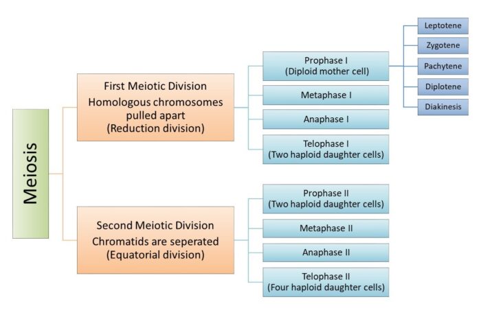
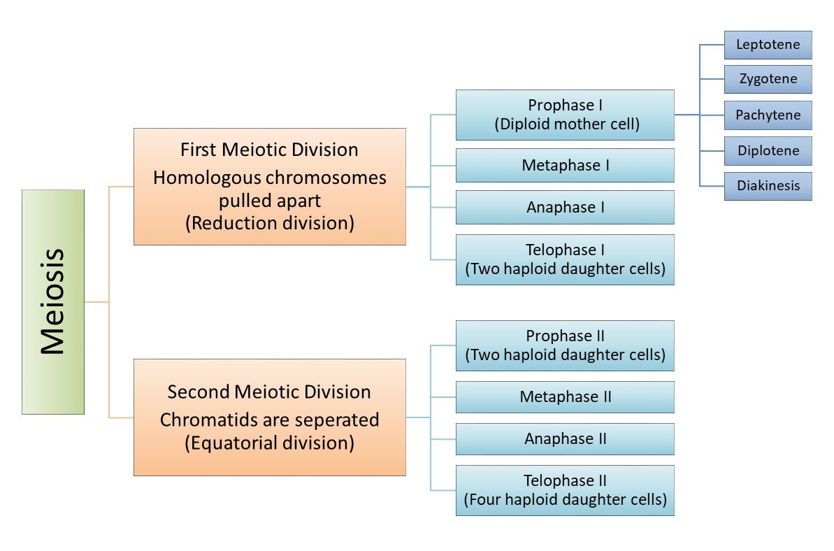
MEIOSIS STAGES
Meiosis divides into two main division
- first meiotic division (meiosis I)
- second meiotic division (meiosis II)
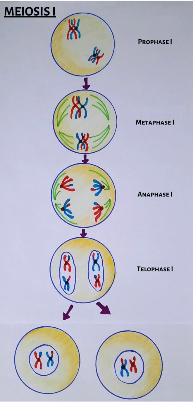
First Meiotic division (meiosis 1)
During meiosis I, homologous chromosomes first pair with one another and then segregate to different daughter cells. Sister chromatids remain together, so completion of meiosis I result in the formation of daughter cells containing a single member of each chromosome pair (consisting of two sister chromatids).
The first meiotic division has four phases namely:
- prophase I
- leptotene
- zygotene
- pachytene
- diplotene
- diakinesis
- metaphase I
- anaphase I
- and, telophase I
Prophase I
Prophase I is quite complex as compared to mitosis. In prophase 1, the events of nucleolar and nuclear envelope disaggregation and intricate chromosome coiling occur.
Typically, prophase I is the longest phase as compared to mitotic prophase. In human females, for example, oocytes in ovary enter prophase I at the time of birth and remain in the same stage of meiotic prophase for several years (since a female produces no additional oocytes in her lifetime). During menstruation, an oocyte matures and released at each cycle.
Since this phase is quite long and complex, it is divided into five sub-stages. These are as follows:
- leptotene
- zygotene
- pachytene
- diplotene
- diakinesis
Leptotene
It is the first sub-stage of prophase I. It consists of the condensing of the already replicated chromosomes.
The already duplicated chromosomes appear as single threads with bead-like thickenings termed chromomeres. The chromosomes at this stage show a definite orientation and a tendency to be polarized. This peculiar arrangement is called the bouquet stage. This is a very short stage of Prophase 1. After a few hours, the nucleus enters the zygotene stage.
Zygotene
During this stage, the paternal and maternal homologous chromosome begins to attract and joined to each other to forms a pair of bivalents. This event constitutes a process called synapsis. This synapsis can form up and down the chromosomes allowing numerous points of contact called the synaptonemal complex.
The synaptonemal complex is a key structure in the lining up of homologous chromosomes. Once formed, the complex is required for continued pairing and chiasma formation during pachytene.
Pachytene
The end of synapsis marks the end of zygotene and beginning of pachytene. Pachytene may last for up to several weeks. During pachytene, chromosomes split longitudinally to form two sister chromatids. The two sister chromatids separate from each other, but the homologous chromosomes remain attached. This makes the complex look much thicker.
At this stage, the nucleoli are clearly visible. The significant events in this stage are the exchange of paternal and maternal genes and chiasma formation.
The synaptonemal complex gradually disappears late in this phase. Recombination between homologous chromosomes will complete by the end of pachytene.
Diplotene (longest phase)
The synaptonemal complex disappears at the diplotene stage and the homologous chromosomes separate along their length. However, the centromere will not split and the longitudinal separation of the homologous chromosome is incomplete. At this stage, the paired chromosome appear to repel each other but attached at few sites along with the pair. These sites refer to as chiasmata. The chiasmata are generally located at the sites on the chromosomes at which genetic exchange during crossing over had previously occurred.
Also, there may be one to several chiasmata in each pair of homologous chromosome.
During diplotene, chromosomes condense so that they appear short and thick by the next stage.
Diakinesis
Diakinesis represents the transition to metaphase during which the meiotic spindle assemble and the chromosomes will prepare for separation. It is the final stage of prophase I and is the termination of the condensing chromosomes. During this stage, the nucleolus and nuclear envelope disappear and the spindle begins to form.
Diakinesis ends with the disappearance of the nucleolus, the breakdown of the nuclear envelope, and the movement of the tetrads to the metaphase plate.
key points in prophase 1
- Homologous chromosomes forms bivalents.
- chiasmata forms between non-sister chromosomes and crossing over occurs.
- exchange of genetic material takes place between non-sister chromatids.
Metaphase I
As the prophase I of meiosis completes, the cell enters metaphase I. The kinetochores of sister chromatids are adjacent to each other and oriented in the same direction. At this stage, the tetrads move towards the centre of the cell and arrange themselves to the metaphase plate.
The orientation of the maternal and paternal chromosomes of each bivalent on the metaphase I plate is random.
Key points
- homologous chromosomes arrange at metaphase plate.
- orientation of chromosomes of each bivalent is random.
Anaphase I
Anaphase I completes the separation of the homologous pair. The tetrads separate so that each homologous double-chromatid chromosome moves to one of the daughter cells. Please note that only homologous chromosomes will separate, not the sister chromatids.
Separation of homologous chromosomes at anaphase I require the dissolution of the chiasmata that hold the bivalents together.
Key points
- Homologous chromosomes will separate and moves to one of the daughter cells (opposite poles).
- chiasmata dissolve.
Telophase I
This is the reduction division step and results in two daughter cells. During telophase I, the chromosomes unfold and the nuclear membrane and nucleoli form. This is followed by meiosis II.
At the completion of meiosis l, each daughter cell has therefore acquired one member of each homologous pair, consisting of two sister chromatids. These are therefore considered haploid cells. These cells take a short rest before entering the second division of meiosis, meiosis II.
Key points
- nuclear envelope will form.
- pair of chromosomes arrives at the poles.
Meiosis II (second meiotic division)
After meiosis I, the cell enters into the stage called interkinesis. It is a short-lived stage. Meiosis II resembles a normal mitosis. Like meiosis I, it is further divided into the following stages:
- Prophase II
- Metaphase II
- Anaphase II
- Telophase II
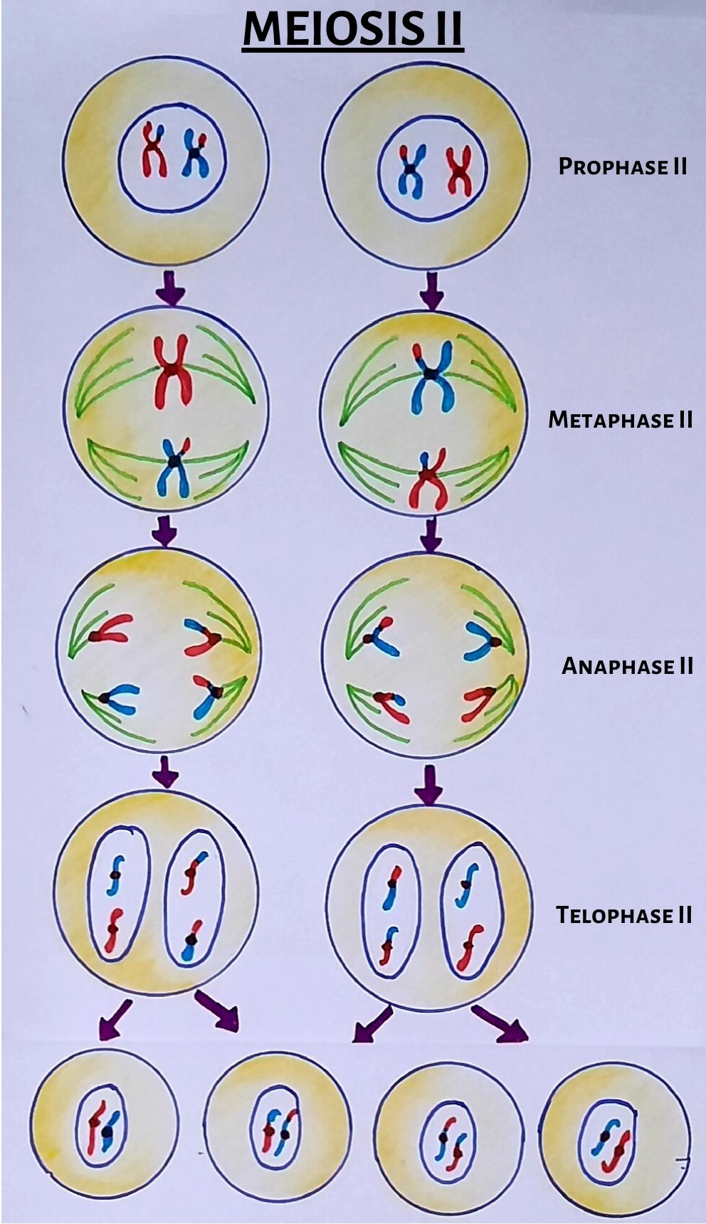
Prophase II
Prophase II is similar to prophase I. If the nuclear envelope had reformed in telophase I, it is broken down again in prophase II. Further, the chromosomes begin to get pulled toward the metaphase plate.
Metaphase II
Unlike metaphase I, the kinetochores of sister chromatids of metaphase II face opposite poles and become attached to opposing sets of chromosomal spindle fibers.
Anaphase II
Anaphase II begins with the synchronous splitting of the centromeres, which had held the sister chromatids together, allowing them to move toward opposite poles of the cell. Basically, this separation marks the final division of the DNA.
Telophase II
It is the final stage of meiosis II in which the chromosomes are once again enclosed by a nuclear envelope.
The final product of meiosis is a true haploid cell with respect to both chromosome number and DNA content. Thus, four cells containing a haploid set of chromosomes will produce at the end of meiotic cell division.
SOURCES AND LINKS
Cell biology: organelle structure and finction., page no. 484-491.
Molecular Biology of the cell, 5th edition, Bruce Alberts, Alexander Johnson, Julian Lewis, Martin Raff, Keith Roberts, and Peter Walter, chapter no. 17.
Karp’s cell and molecular biology: concepts and experiments 8th edition by Janet Iwasa and Wallace Marshall, chapter 14.
The cell: A molecular approach by Geoffrey M Cooper, chapter 16, the cell cycle.
Cell and Molecular biology by Prakash S. Lohar, page no. 209-204.