WHAT ARE GRAM NEGATIVE BACTERIA?
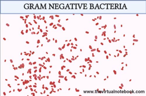
Gram-negative bacteria are bacteria that do not retain crystal violet dye in the Gram staining protocol due to a thinner cell wall. They appear red or pink in colour due to the counterstain (commonly safranin). The Gram staining technique is developed by Hans Christian Gram hence, named after him.
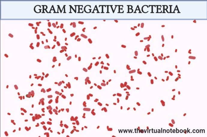
In gram staining technique, the gram-negative bacteria do not retain the ‘dye-iodine complex’ and become translucent after a brief wash with alcohol or acetone while the Gram-positive bacteria retain the ‘dye-iodine complex’ (purple colour). In the next step, culture can be counterstained with safranin (a red dye). Thus, gram-positive bacteria look purple under the microscope whereas gram-negative bacteria look red or pink.
Surprisingly, Gram-negative bacteria are present in all environment that support life. And do you know that almost most of the gram-negative bacteria cause diseases and some of them are life-threatening? And the worst part, they are resistant to multiple drugs, therefore, increasingly resistant to most available antibiotics.
Let’s move on to the most important components of gram-negative bacteria i.e., the cell wall…
CELL WALL OF GRAM-NEGATIVE BACTERIA
The cell wall is the outermost boundary of the bacterial cell. The bacterial cell wall can withstand and maintain the osmotic pressure gradient between the interior and exterior cellular environment and give the cell its outer form. It also facilitates communication with its outer surrounding.
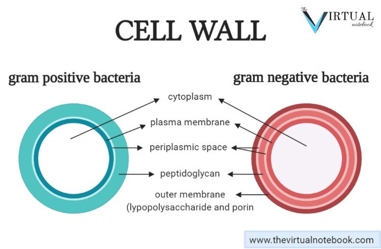
It is evident that gram-negative cell walls are much more complex than gram-positive walls. The thin peptidoglycan layer next to the plasma membrane and bounded on either side by the periplasmic space usually constitutes only 5 to 10% of the wall weight.
Components
The architecture of Gram-negative bacteria is fundamentally different from that of Gram-positive bacteria. The amount of peptidoglycan is lesser and it forms a single-layered sheet around the cell.
1. Periplasm
It is an intermembrane structure, lying between the cell membrane and the outer membrane. In addition, it ranges in width from 1 nm to as great as 71 nm. External to this periplasm is an elaborate outer membrane.
2. Outer membrane
The outer membrane of Gram-negative bacteria is biologically unique. Its inner leaflet consists of ordinary phospholipids, but these are replaced in the outer leaflet by a special molecule called lipopolysaccharide (LPS), which is extremely toxic to humans and other animals, and thus commonly called endotoxin. Even in minute amounts, these endotoxins are so dangerous that they can cause a fever and shock syndrome called gram-negative or endotoxic shock during the course of a gram-negative infection.
3. Lipopolysaccharides(LPS)
As mentioned earlier, it is extremely toxic to humans and other animals. These large, complex molecules contain both lipid and carbohydrate, and consist of three parts:
- Lipid A: contains two glucosamine sugar derivatives, each with three fatty acids and phosphate or pyrophosphate attached.
- A core polysaccharide: joined to lipid A and contain some unusual carbohydrate residues and fairly constant in structure among related species of bacteria.
- O antigen polysaccharide side chains: constitutes the major surface antigen of gram-negative cells. It has several peculiar sugars and varies in composition between bacterial strains.
Functions of lipopolysaccharide (LPS)
- Its major function is to help in creating a permeability barrier.
- It contributes to the negative charge on the bacterial surface.
- Also, it helps in stabilizing the outer membrane structure.
4. Porins
The presence of the outer membrane results in the covering of gram-negative cells that creates a formidable permeability barrier. This barrier is overcome by special structure proteins called porins. Because they create pores through the outer membrane, makes it possible for hydrophilic solute molecules to diffuse through it and into the periplasm.
CHARACTERISTICS OF GRAM-NEGATIVE BACTERIA
These are the following characteristics of gram-negative bacteria:
- An inner cell membrane is present (cytoplasmic membrane).
- The thinner peptidoglycan layer is present in this class of bacteria.
- Periplasmic space is present between the layers of peptidoglycan and the secondary cell membrane.
- Porins are present on the outer membrane, which act like pores for particular molecules.
- If present, flagella have four supporting rings instead of two.
- Most of the Gram-negative bacteria do not form spores. Though, there are some exceptions.
- Lipoproteins are present in the cell wall.
EXAMPLES
- Protobacteria: they include a wide variety of pathogens, such as Escherichia, Salmonella, Vibrio, Helicobacter, and many other notable genera. Others are free-living and include many of the bacteria responsible for nitrogen fixation.
- Enterobacteriaceae: it is a large, heterogeneous family of gram-negative bacteria and several members live in the intestines of animals. Examples:- Enterobacter, Citrobacter, Moraxella, Pseudomonas, helicobacter, etc.
- Acetic acid bacteria: they oxidize sugars or ethanol and produce acetic acid during fermentation.
- Cyanobacteria: these are free-living photosynthetic bacteria and often called blue-green algae. Certain types (five types) of cyanobacteria produce toxins namely hepatotoxins, neurotoxins, and, lipopolysaccharides endotoxin.
- Spirochetes: they are free-living, anaerobic (with some exceptions), and spirally-coiled bacteria that can cause diseases in humans such as syphilis and Lyme diseases. Examples of genera of spirochetes include Spirochaeta, Treponema, Borrelia, and Leptospira.
- Green sulfur bacteria: they are strictly anaerobic, photosynthetic, aquatic bacteria. They occur in aquatic sediments, sulfur springs, and hot springs and utilize reduced sulfur compounds instead of oxygen. Additionally, they use reduced sulfur compounds as their electron donors and fix carbon using the reverse TCA cycle.
- Green non-sulfur bacteria: they utilize light as their energy source and usually present in photosynthetic bodies called chlorosomes.
DISEASES CAUSED BY GRAM-NEGATIVE BACTERIA
Gram-negative bacteria cause so many diseases in humans and some are listed below:
Sexually transmitted diseases (STDs)
STDs can spread from one person to another, usually during vaginal, anal, and oral sex. Sometimes, these infections can be transmitted nonsexually, such as from mother to infant during pregnancy or childbirth, or through blood transfusions or shared needles. For example, some sexually transmitted diseases are as follows:
- Syphilis: caused by Gram-negative, spirochetal bacterium T. pallidum.
- Gonococcal infection: caused by the Gram-negative diplococci N. gonorrhoea.
- Chlamydiosis:caused by the bacterium Chlamydia trachomatis.
Respiratory diseases
Respiratory tract infections are the most frequently reported infections of the human being. There are four medically important gram-negative rods typically associated with the respiratory tract, namely,
- Haemophilus influenzae: It is an important cause of upper respiratory tract infections such as otitis media, sinusitis, conjunctivitis, and epiglottitis. Also, it causes sepsis in children and pneumonia in adults, particularly in those with chronic obstructive lung disease.
- Bordetella pertussis: it causes Pertussis. It is a very contagious respiratory disease commonly known as whooping cough.
- Legionella pneumophila: it causes Legionnaires disease (a severe form of pneumonia).
- Acinetobacter baumannii: it causes hospital-acquired pneumonia (HAP).
Urinary diseases
A urinary tract infection (UTI) is an infection in any part of the urinary system (kidneys, ureters, bladder and urethra). Most infections involve the lower urinary tract, i.e., the bladder and the urethra. An interesting point to know is that women are at greater risk of developing a UTI than men (why women suffer more?…. huh… frustrating).
Causative agent
- E.coli: it can cause infection of the bladder, i.e., cystitis.
- Proteus mirabilis: it is capable of causing symptomatic infections of the urinary tract including cystitis and pyelonephritis.
- Enterobacter cloacae: can cause UTIs including Bacteremia.
- Serratia marcescens: responsible for nosocomial urinary tract infections (UTIs).
Gastrointestinal diseases
Gastrointestinal diseases refer to diseases involving the gastrointestinal tract, namely the oesophagus, stomach, small intestine, large intestine and rectum, and the accessory organs of digestion, the liver, gallbladder, and pancreas.
Causative agent
- Helicobacter pylori: responsible for a number of digestive tract disorders, such as chronic active gastritis, peptic ulceration, gastric cancer, and mucosa-associated lymphoid tissue lymphoma.
- Salmonella enteritidis: humans infected with Salmonella may develop diarrhoea, fever and abdominal cramps.
- Salmonella typhi: can cause local intestinal inflammation and diarrhoea.
TREATMENT
One of the several unique characteristics of Gram-negative bacteria is the structure of the outer membrane. It comprises a complex lipopolysaccharide whose lipid portion acts as an endotoxin. If endotoxin enters the circulatory system, it causes a toxic reaction, with the sufferer developing high temperature, high respiration rate, and low blood pressure. This may lead to endotoxic shock, which may be fatal.
This outer membrane protects the bacteria from several antibiotics, dyes, and detergents that would normally damage the inner membrane or cell wall (peptidoglycan). In addition, the outer membrane provides these bacteria with resistance to lysozyme and penicillin. However, alternative treatments such as lysozyme with EDTA and the antibiotic ampicillin have developed to combat the protective outer membrane of some pathogenic Gram-negative organisms. Other drugs can also be used, significant ones being chloramphenicol, streptomycin, and nalidixic acid.
GRAM-NEGATIVE VS GRAM-POSITIVE BACTERIA
- Cell envelope: the very first difference between Gram-positive and Gram-negative bacteria lies in the thickness of the cell wall. The cell envelope of gram-positive organisms consists of a thicker cell wall as compared to Gram-positive bacteria.
- Lipid membrane: Gram-positive bacteria have no outer lipid membrane whereas Gram-negative bacteria have an outer lipid membrane.
- Periplasmic space: the periplasm is an intermembrane structure, lying between the cell membrane and the outer membrane(unique to gram-negative cells). This space is present in Gram-negative bacteria while sometimes absent in Gram-positive bacteria.
- Peptidoglycan layer: In Gram-positive bacteria, the peptidoglycan layer of the cell wall is thick. However, it is thinner in Gram-negative bacteria.
- Resistance to osmotic pressure: because of the thicker peptidoglycan layer, the walls of gram-positive cells are more resistant to osmotic pressure than those of gram-negative bacteria.
- Staining dye used: crystal violet dye(purple or blue) is used to identify Gram-positive bacteria. On the other hand, safranin is used to identify the Gram-negative bacteria which gives it a pink or red colour.
- Resistance to antibiotics: Gram-negative bacteria are more resistant to antibiotics as compared to Gram-negative bacteria. This resistance ability of Gram-negative bacteria makes them more dangerous.
SOURCES AND EXTERNAL LINKS
Sherris Medical Microbiology by Kenneth J Ryan, chapter no. 21: Bacteria-basic concepts
Prescott’s Principles of Microbiology by Willey, Sherwood and Woolverton chapter no. 3: Prokaryotic cell structure and function
Jawetz, Melnick, & Adelberg’s Medical Microbiology; Twenty-Sixth Edition., chapter no. 2 and 3-page no. 39,48,49.
https://en.wikipedia.org/wiki/Acetic_acid_bacteria
https://www.sciencedirect.com/topics/agricultural-and-biological-sciences/cyanobacteria
https://www.dictionary.com/browse/green-sulfur-bacteria
https://accessmedicine.mhmedical.com/content.aspx?bookid=2381§ionid=187689313
https://www.technologynetworks.com/immunology/articles/gram-positive-vs-gram-negative-323007