PLANT CELL DEFINITION
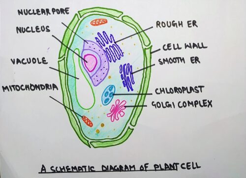
A Cell represents the smallest structural and functional unit of life. Plants are multicellular eukaryotic organisms and belong to the kingdom Plantae. Plant cells have a membrane-bound nucleus. In addition, cells vary greatly in size, form, structure, and function. Some are measured in micrometres, others in millimetres, and still others in centimetres (fibres in certain plants). Although the plant cell structure is very similar to that of the animal cell, the two types of cells contain many of the same organelles. These organelles carry out the same functions in both types of cells. Aside from common organelles, plant and animal cells are very different. Plant cells are very unique because of the presence of three additional structures. These structures (cell wall, vacuoles, and plastids) are important to a plant’s ability to function.
If I assume that I know nothing about a plant cell and it’s totally an alien term for me, what or which questions come into my mind? Firstly, I will ask, what is a plant cell? What purpose does it serve? How does it look like? etc., etc., etc.,… Questions can be countless. I tried to answer all these questions in this article.
Plant Cell Structure
They vary considerably in size, shape and structure. It is, therefore, difficult to present a general picture of plant cells. Some are biochemically inactive, such as those in the xylem. Also, cells of the phloem lack the nucleus and are not regarded as fully active.
The connection between adjacent cells through plasmodesmata is an important feature of plant cells. Typically, plant cells are cubic or rectangular in shape and a lot bigger than animal cells in structure. Comparatively, each cell can contain one large vacuole or several smaller vacuoles. The cytoplasm is an aqueous medium of different viscosity and composition which contains membrane-bound bodies known as organelles. Also embedded in the cytosol are chloroplasts (plastid), mitochondria, ribosomes, Golgi apparatus, peroxisomes, glyoxysomes, spherosomes, microtubules and microfilaments (part of the cytoskeleton).
Plant cell organelles
As stated above, a plant cell has various organelles:
- Cell wall
- Cytoplasm
- Plasma Membrane
- Nucleus
- Ribosomes
- cytoskeleton (microtubules, microfilaments, intermediate filaments)
- Golgi bodies/Golgi complex
- Endoplasmic reticulum
- Plastids
- mitochondria
- Vacuoles
1. The cell wall
Plant cell wall definition
It is the outermost boundary of the eukaryotic plant cell. Rapidly growing cells (e.g. meristematic tissue) are surrounded by thin primary cell walls. Once growth ceased, the secondary wall will form between the primary wall and the cell membrane.
The plant cell wall is mainly composed of cellulose, hemicellulose and pectin
The primary cell wall is 1-3µm thick and begins to form between dividing cells. The plant cell wall is mainly composed of cellulose, hemicellulose and pectin. In addition, Extensin and Arabinose are the major structural protein of the cell wall. Although, Cellulose is the main component of the cell wall and is insoluble in water. It is stable at low pH (dilute acids) and has a flattened ribbon-like shape. The amorphous wall matrix consists mainly of hemicelluloses and pectic polysaccharides. Pectins are the most water-soluble component of the cell wall.
Plant cell wall structure
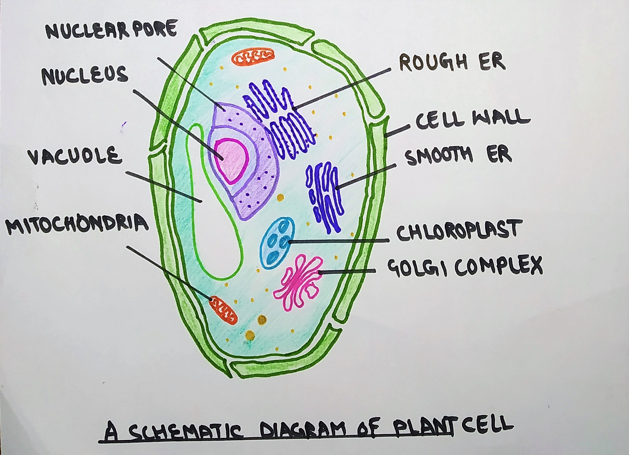
A typical plant cell has three distinct regions:
- Primary wall (thin and elastic)
- Secondary wall (thick and rigid)
- Middle lamella
The primary wall is present in growing cells (meristematic tissues in case of the plant cell), comprises 30-40% cellulose(dry weight), 50% of polysaccharides ( hemicellulose & pectin) and 5% glycoproteins rich in hydroxy proteins.
The secondary wall is generally very rigid and does not alter its shape. Generally, the protoplasts of the primary wall secrete the secondary wall materials when the cells have stopped enlarging. The protoplast then totally diminishes and only the wall remains.
Middle lamella is present between the two adjacent cell walls. In other words, it is sandwiched between the primary and secondary walls. This middle lamella comprises pectin, cellulose, calcium, magnesium pectate, etc.,
Functions of plant cell wall
- The Cell wall is rigid which means ‘it provides shape to the plant cell’.
- It acts as a physical barrier protecting cells from invading viruses, fungi, bacteria, etc. Thus, we can say that they serve as a part of a defence mechanism.
- It presents a barrier to cell expansion during cell division.
- It is semipermeable hence it allows the circulation of materials such as water, molecular nutrients, and minerals.
2. Cytoplasm
The term cytoplasm was originally introduced by Kölliker (1862), used to designate all of the material surrounding the nucleus. It is a gel-like colloidal material lying just below the cell membrane. It includes several substances either in dissolved or in a suspended condition. Provided that it is not classified as one of the cell’s organelles because it doesn’t possess major roles except being a physical medium for holding and housing most of the cell organelles.
Structure of cytoplasm
The cytoplasm contains about 90% of water and the remaining 10% are organic and inorganic substances. The organic substances include carbohydrates, proteins amino acids, glycerol, hormones, vitamins and nucleic acids.
3. Plasma membrane
It is a membrane that separates the interior of the cell from the outside environment. A cell wall is attached to the plasma membrane on its outside surface and the interior to the cell is cytoplasm. Among the various membranes of the cell, the plasma membrane typically has the clearest dark-light-dark or unit membrane appearance in electron micrographs.
Several cell biologists attempted to understand the molecular structure of the cell membrane and proposed different models for the same. These models are classified into two basic categories:-
- Bilayer model
- Micellar model
In the bilayer model, the proteins and lipids are arranged in the form of layers whereas there are small and similar independent subunits present in the plasma membrane. Examples:-unit membrane model, Daniell-Davson model.
Functions
- It provides a framework in which components can be organized.
- It acts as a selectively permeable membrane. The Plasma membrane allows the cell to exchange material and information.
- It also acts as a site where energy is transduced (convert) from one type to another.
- Membranes also provide mechanical strength and electrical insulation.
4. Nucleus
The nucleus contains the blueprint for determining the structure and function of cells. It is the largest organelle present in the cytoplasm. It is about 10µm in diameter in non-dividing cells.
Structure of Nucleus
A typical nucleus shows the following components:
- Nuclear envelope
- Nuclear sap
- Chromatin
- Nucleolus
4.1 Nuclear envelope
It is a double-membrane structure that is 10-50nm in thickness. Basically, the inner & outer membrane of the nuclear envelope has a thickness of about 70-80Å and is mainly composed of proteins & lipids. The outer membrane is usually studded with ribosomes and is often seen continuous with the membrane of rough ER. Conversely, the inner membrane is lined by a dense, fibrillar network, known as nuclear lamina of varying thickness. This nuclear lamina provides structural support to the nuclear envelope.
Double membranes fuse with each other at irregular intervals leading to the formation of nuclear pores. Each nuclear pore has a diameter of about 60-90nm. In addition, the nuclear pore contains a complex, basket-like apparatus called the nuclear core complex. This complex fills the
4.2. Nuclear sap
Nuclear sap is a semi-fluid, gel-like substance present in the nucleus. It is also known as nuclear matrix, karyoplasm or nucleoplasm (just like the cytoplasm present inside a cell, the nucleus contains nucleoplasm). It contains nucleoproteins, nucleic acid, enzymes, and minerals.
4.3. Chromatin
Chromatin is a complex of macromolecules comprises of DNA, RNA, and protein. It is present inside the nucleus of eukaryotic cells.
The key protein in chromatin is the histones (a
Histones help DNA to organize into bead-like structures called nucleosomes. The amount of DNA tightly folded with each nucleosome bead is approx. 200bp. However, the no. of bp associated with each bead varies from about 150-250.
In addition, a nucleosome is wrapped around a set of 8 histones called an octamer. The nucleosome can be further folded to produce chromatin fibre. Chromatin fibres are coiled and condensed to form chromosomes.
4.4 Nucleolus
It appears as a spheroid body within the nucleus of eukaryotic cells. Its size can vary from 1 to 5mm approx. Also, the number of nucleoli per nucleus is quite variable.
Functions of nucleus
- The nucleus has immense importance in the storage and utilization of genetic information. It is the repository of most of the cell’s genetic information, passing it on to the daughter cells in the course of cell division.
- Nuclear envelope of nucleus regulates the exchange of electron, ions and molecules of various compounds.
- It controls the ongoing activities of the cell by determining which RNA and protein molecules are produced by the cell and when they are produced.
5. Ribosomes
Ribosomes are ribonucleoprotein particles present in a variety of cells either in a free state or attached to the ER. Their number is especially large in cells that perform active synthesis. Although the number of protein molecules in ribosomes greatly exceeds the number of RNA molecules, RNA constitutes about 60% of the mass of a ribosome.
Structure
Each ribosome
The small subunit has a head and a body separated by a constriction. Although, the body occupies about two-thirds of the volume of this subunit.
Function
Ribosomes are the protein builders or protein synthesizers of the cell.
6. Cytoskeleton
Like we have the skeleton system for support. Similarly, the cell has a cytoskeleton. The cytoskeleton is a network of interconnected protein filaments and tubules that extends from the nucleus to the plasma membrane in eukaryotic cells. Much as bones and muscles give an animal structure and produce movement, the elements of the cytoskeleton maintain cell shape and, along with motor proteins, allow the cell and its organelles to move. But unlike an animal’s skeleton, the cytoskeleton is highly dynamic—its elements can be quickly assembled and disassembled as appropriate. The cytoskeleton includes:-
- Microtubules (actin filaments),
- Microfilaments, and
- Intermediate filaments.
1. Microfilaments (actin filaments)
Microfilaments are the narrowest among these three types. It is 1-2 µm in length and 5-7µm in diameter. Microfilaments of higher eukaryotes are made up of many linked monomers of a protein called actin. Because they are made up of actin monomers, they are also known as actin filaments. Actin filament forms a double helix, 6 nm wide, with globular subunits 5.5 nm long and a 37 nm helix repeat. Like microtubules, microfilaments also have polarity.
Function
- Actin filaments are involved in localized cell membrane movement.
- Another example of actin is the involvement of the sperm of an animal. When the sperm encounters the outer jelly coat of the egg, Ca2+ enters the sperm, causing the acrosome membrane to fuse with the plasma membrane. As a result, the coat is dissolved and the plasma membrane of sperm and the egg can fuse to form a zygote.
- Microfilament also mediates the cytoplasm streaming or cyclosis in most of the cells.
2. Intermediate filament
These are smooth surfaced, solid and unbranched filaments having a diameter of 7-11 nm that lies between those of microtubules and microfilaments. Unlike microfilament and microtubules, intermediate filaments are chemically heterogeneous in structure, composition, and solubility.
The basic unit of intermediate filament is the ‘two-chain coil’ which are identical ( homopolymer) and are formed from helix interactions. On the other hand, cytokeratin is heterodimers, with one chain of one type and the other of a different type. Once the dimer has formed, it interacts with a second dimer to form a tetramer(in parallel fashion), two of these units then associate in anti-parallel fashion to form a protofilament. Now, four protofilaments make up the 10 nm diameter filament seen in cells.
Function
Intermediate filaments play a significant role as motility molecules within the cytoplasm of different classes of cells.
- The keratin filaments of epithelial cells form a network around the nucleus and also ramify through the cytoplasm where their role is to hold the organelles into place.
- Desmin ( in skeletal muscles) filament forms an organized network at the Z-line where they help to integrate muscle function.
- Neurofilaments play an integral part in axon transport of different macromolecules including synaptic protein towards the presynaptic membrane.
- Intermediate filaments provide the clues to the clinicians with cell markers to determine the identity of the tumor.
3. Microtubules
In 1800, microscopists have reported that cells and their extensions often contain a fibrous component. This was especially noticeable in cilia and flagella. In 1963, D. Slautterback coined the term microtubules. These are present virtually in all eukaryotic cells.
Structure
In animal cells, they usually radiate from a centrosome located near the nucleus while in plant cells they are more near the plasma membrane. They appear in longitudinal sections as long, thin rods or in cross-sections as hollow tubes ringed by 13 subunits. The outer and inner diameter of these tubules is 30nm and 14nm respectively. Therefore, the wall thickness is 18nm.
A clear, unstained zone, 5-20nm in width, often surrounds the outer wall of the microtubule. The length of microtubules
They comprise polymers of tubulin protein subunits, which are of two types:
- α tubulin molecules
- β tubulin molecules
Microtubules are dynamic structures because they can easily change their length by removing tubulin dimers. This process is controlled by the centrosome which lies near the nucleus. Microtubules radiating from the centrosome help maintain the shape of the cell and act as tracks along which organelles and other materials can move.
Function
- microtubules provide an internal skeleton or scaffold that provides structural support and helps to maintain the position of cytoplasmic organelles.
- They serve as the primary components of the machinery responsible for mitosis and meiosis.
- Microtubules form principal elements of cilia and flagella as motile cell projections.
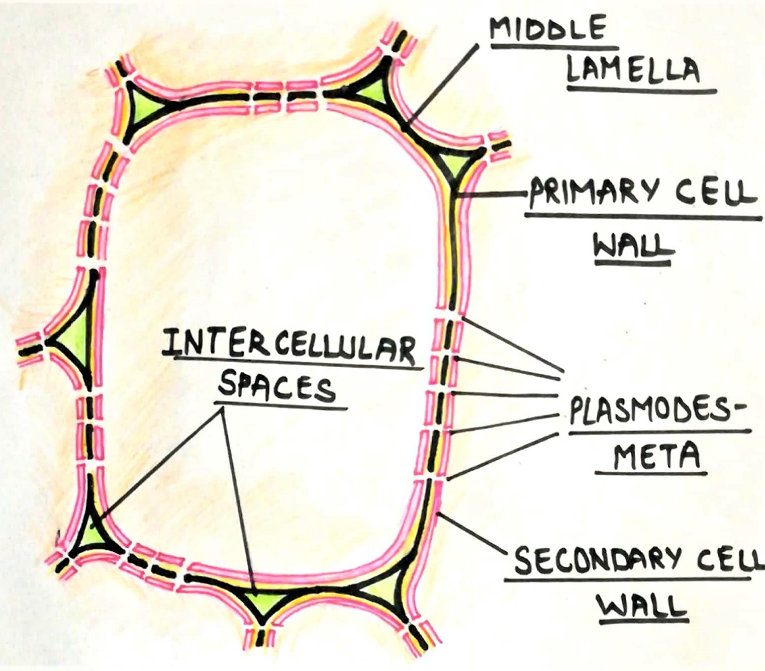
7. Golgi Complex (editor of synthesized protein)
This is a membranous organelle present in the cell’s cytoplasm. You can say that the Golgi complex is an organelle that sorts and modifies proteins & lipids (fats).
Structure
Golgi complex appears as a complex array of interconnected cisternae, tubules & vesicles. It comprises several fattened, stacked sacs referred to as cisternae. In addition, each Golgi body consists of 4-8 cisternae, slightly curved, each having a diameter of 0.5-1.0mm. The membrane of the cisternae is porous. Moreover, the pores may be localized or present all over the surface. Cisternae of the Golgi apparatus has two faces– cis and trans face.
The cis face is convex and closer to the endoplasmic reticulum. It acts as receiving compartment from the endoplasmic reticulum.
On the opposite side is the trans face, which is present towards the plasma membrane. In between cis and trans cisternae, the stack of cisternae known as medial cisternae.
For the role in protein secretion, see Golgi complex and its role in protein secretion
Functions
- Golgi complex is the site of synthesis and concentration of glycoproteins and mucopolysaccharides.
- In the plant cells, it plays an indispensable role in the formation of the cell plate during mitosis.
- It associates with the formation of melanin in animal cells.
- Golgi complex is responsible for the modification of non-secreted proteins.
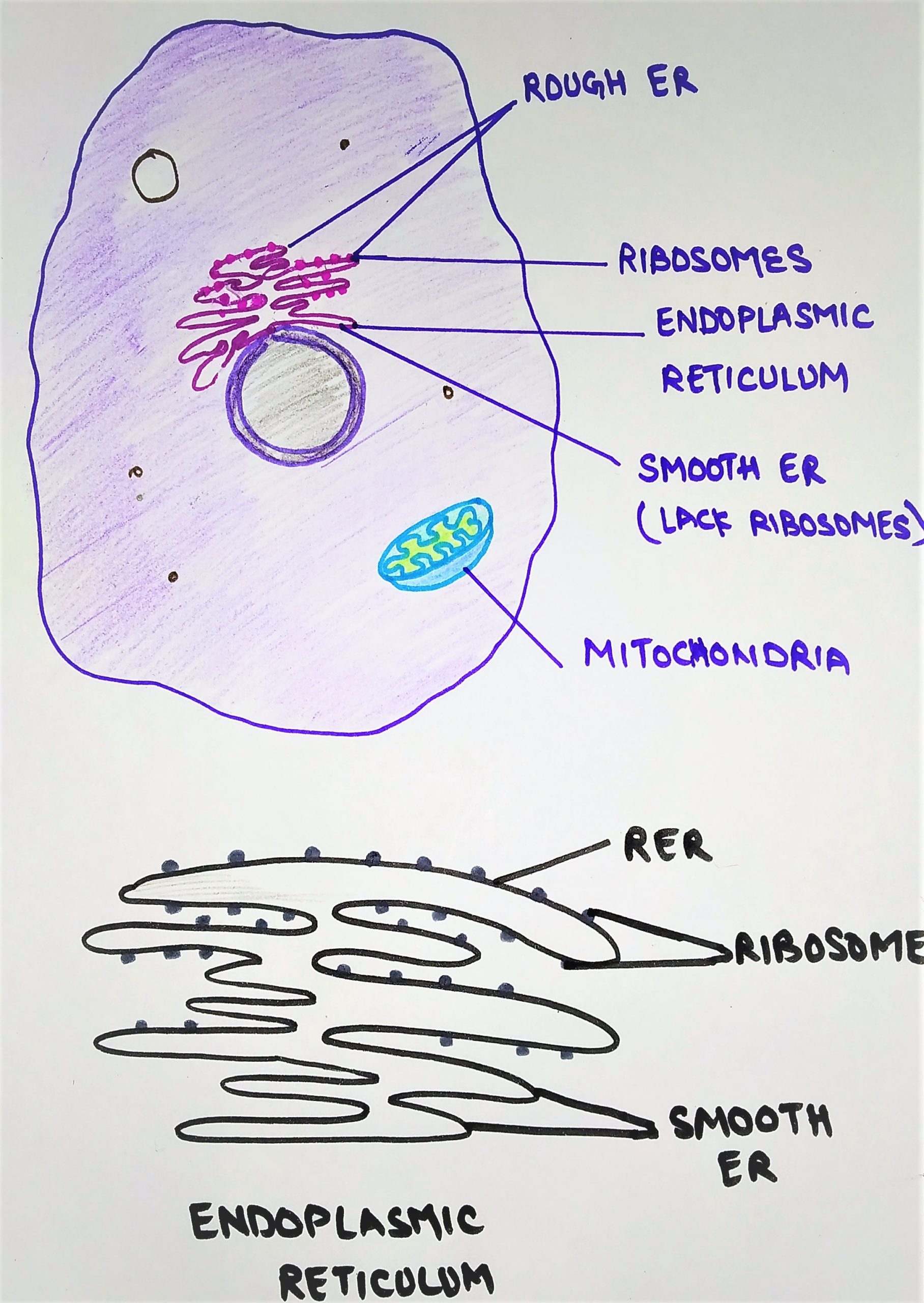
8. Endoplasmic Reticulum
The Endoplasmic reticulum consists of a system of interconnected tubules with the membrane surrounding an interior cisternal space.
The Endoplasmic reticulum membrane is generally thinner than the
Typically, the cisternal space is 30nm in diameter but can increase considerably when induced by drugs. Due to its huge surface area, it is the largest membrane in the cell.
The outstanding morphological difference is the presence/ absence of ribosomes that are present/absent on their outer surface. Depending upon the presence/absence of ribosomes, it is of two types:-
- Rough ER (rough endoplasmic reticulum)
- smooth ER (smooth endoplasmic reticulum)
Rough ER
It is studded with numerous 18-22
Smooth ER
They lack ribosomes on their outer membrane. They appear to be more vascular with swollen cisternae. It occurs along with RER in some cell types, such as liver hepatocytes, but is more commonly proliferated in specialized cells lacking RER.
Functions
- It plays a major role in the synthesis of the secretory proteins and their modifications.
- ER plays an indispensable role in the synthesis of glycogen, lipids, cholesterol and steroid hormones.
- It helps in the formation of a nuclear envelope and other cell organelles.
- It plays a major role in detoxification of certain drugs and muscle contractions.
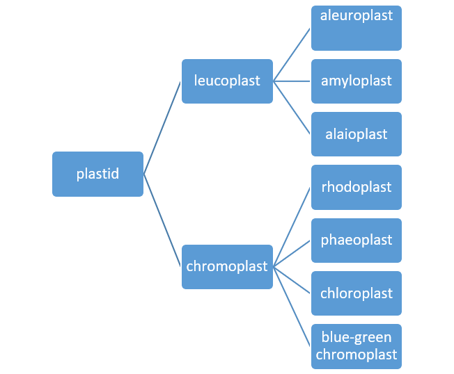
9. Plastids
These are double membrane-bound organelles present in most of the plant cells and some protozoans. They are generally absent in animal cells, bacteria, and fungi. Plastids
They are of two types:
- Leucoplast (colorless)
- Chromoplasts (color)
Leucoplast:- they generally serve the purpose of storage. These are generally present in cells that do not receive direct sunlight. Leucoplasts are of three types:
- Aleuroplasts (store proteins)
- Amyloplast (store starch)
- Elaioplast ( store oil)
Chromoplast:- they are colour plastids of various colour pigments. They act as a primary site for trapping and converting solar energy.
Based on their colored pigment, chromoplast are of following types:-
- Chloroplast (green)
- Phaeoplast ( brown)
- Rhodoplast (red)
- Blue-green chromoplast ( blue green)
Structure
Chloroplasts show great variation in shape, size, and numbers. Either they may have oval, disc-shaped or spherical structure or it may be band-shaped, ribbon-shaped.
Chloroplasts have a double-membrane envelope, 6-8nm thick each (phospholipid layer), consists of the inner and outer membrane. Further, the spaces between the double membranes covered with an aqueous matrix known as stroma having a thickness of about 10-20nm. This aqueous matrix contains various enzymes and proteins that are essential for cellular processes. The chloroplast shows a complicated lamellar system composed of small cylindrical structures called
In 1947, S. Granick and K. porter isolated chloroplast and showed that
The stroma of “matrix” of plastids usually contain several kinds of discrete bodies:-
- Starch grain (2mm in length)
- Postoglobuli (0.1mm in diameter) –a common feature of the chloroplast. Postoglobuli may accumulate colored pigment.
- Ribosomes (17 nm in diameter) –a
universal feature of thestroma . Fibrili (2.5 nm in diameter) – represent chromoplasts DNA.
Functions
- Photosynthesis: major function of the chloroplast is to convert light energy into chemical energy.
- Plastids serve the purpose of the manufacturing and storage of protein, starch, and oil.
- Plastids provide colour to fruits and flowers.
10.Mitochondria
Mitochondria are much smaller than chloroplasts, and they are usually visible only under an electron microscope. They are dynamic organelles that move about the cell and can change their shape. In addition, the mitochondria per cell differ considerably, usually depending upon the metabolic activity of the cell. Moreover, active cells have much more mitochondria than less active ones. Mitochondria are often called the powerhouses of the cell because they produce most of the ATP the cell utilizes.
Structure
They have a sausage-shaped body having a thickness of about 0.5m. Like chloroplasts, mitochondria have double membrane. It is enveloped by two
The outer membrane shows a
The space enclosed by the inner membrane is called the matrix (inner chamber) and is usually filled up by a proteinaceous material. Within the matrix are small granules, small ribosomes, and a double-stranded circular DNA. The inner membrane is thrown into a series of irregular folds, called cristae or cristae mitochondrial. These cristae are tubular or finger-shaped and may be branched.
Function
- The mitochondria transduce chemical energy used by the cell in synthetic reaction and active transport.
- Mitochondria store and transport ATP molecules.
- Mitochondria also involved in the biogenesis of haem.
- The proton gradient set up by electron transport can produce heat in newborns, hibernating animals and cold stressed animals.
11. Vacuoles
Together with the presence of plastids and a cell wall, the vacuole is one of the three characteristics that distinguish plant cells from animal cells. They have a single membrane. Within many cells, up to 90% of the cell volume may be occupied by a single vacuole or multiple vacuoles. In meristematic tissue, several smaller vacuoles (
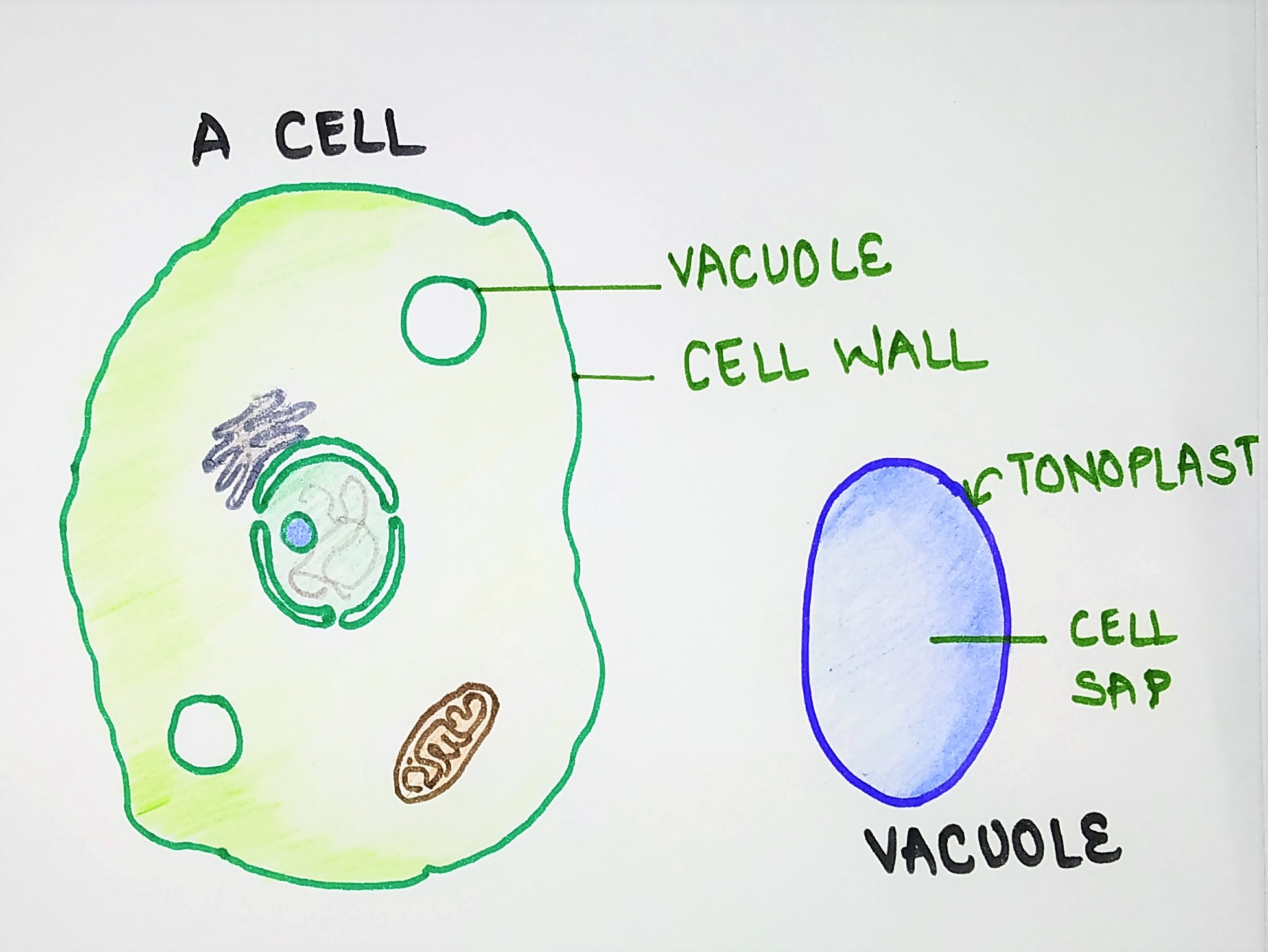
Vacuoles contain solutes in higher concentrations (0.4-0.6 M). For example, citrus fruits store approximately 0.3 M citric acid in vacuoles which can be squeezed out as juice. This compartmentation, therefore, prevents many important enzymes from being denatured.
Functions
- It serves as a storage organelle. Some storage substances are waste products of the cell whereas other stored substances serve as food (energy) reserves.
- Vacuoles are also rich in hydrolytic enzymes and therefore function in a similar manner to animal lysosomes.
For more info on vacuoles, see: vacuoles and microbodies
TYPES OF PLANT CELLS
plant cell differentiate from undifferentiated meristematic cells to form major classes of cells and tissues. These are as follows:
- Parenchyma
- collenchyma
- sclerenchyma
- xylem
- phloem
- epidermis
1. Parenchyma
These are live undifferentiated cells that have functions ranging from storage, support, photosynthesis, and secretion of waste materials. They are green in colour and leaves comprises mainly of parenchyma cells. In addition, parenchyma cells have thin, permeable primary walls enabling the transport of small molecules between them, and their cytoplasm is responsible for a wide range of biochemical functions. These cells are typically more flexible than others because they are thinner.
2. Collenchyma
They are elongated, hard or rigid cells present below the epidermis. They are also present in young plants on the outer layers of their stems and leaves. collenchyma cells are alive at maturity and have thickened cellulosic cell walls. During maturity, at some point, they resemble the parenchyma cells which transform into the collenchyma cells. When a few cells accumulate, the Golgi bodies along with the endoplasmic reticulum come up together to form the primary cell wall. When two cells fuse, they form a thin primary wall that doesn’t differentiate to collenchyma cells.
The role of collenchyma is to support the plant in axes still growing in length, and to confer flexibility and tensile strength on tissues.
3. Sclerenchyma
These cells are more rigid compared to collenchyma cells. Sclerenchyma tissues comprises of two types of cells:
- Sclereids: they are hard, tough cells that give leaves or fruits a gritty texture.
- fibres: they are elongated cells with lignified secondary walls that provide load-bearing support and tensile strength to the leaves and stems of herbaceous plants.
Due to their thickened cell wall, they offer protection and support to other plants’ tissues especially the tree trunks and fibers of large herbal trees.
4. Xylem
Xylem is a complex vascular tissue and composed of two elements for water-conduction: Tracheids and vessel elements. Tracheids are pointed, elongated cells with lignified secondary thickening of the cell walls, specialised for conduction of water. The vessel elements allow the transport of water. They are hollow, shorter, wider than the tracheids but lack the angeled endplates, therefore they are aligned with each other forming a continuous hollow tube, 3 meters long.
In short, xylem cells are the transport cells in vascular plants. They help in the transport of water and minerals from the roots to the leaves and other parts of the plants.
5. Phloem
Phloem cells are present outside the xylem layer of cells. They transport food to different parts of the higher plants. Phloem consists of two main cell types:
- sieve tubes, and
- companion cells.
Sieve tubes are the cells that control the cell’s metabolism, and they are linked together with large numbers of plasmodesmata. The sieve tube elements lack nuclei and ribosomes, and their metabolism and functions are regulated by the adjacent nucleate companion cells.
The companion cells are responsible for loading the phloem with sugars by assisting in moving materials into and out of the sieve tube members.
6. Epidermis/epidermal cells
The plant epidermis/epidermal cells are the external cells of the plants, comprises parenchyma cells, that covers the external surfaces of leaves, stems, and roots. In addition, they offer protection from water loss, pathogenic invaders such as fungi. Three cell types may be present in the epidermis and these are:
- Pavement cells
- Stomatal guard cells
- Trichomes
REFERENCES
Cell biology: organelle structure and function by David E. Sadava. Page no.241-263, page no. 295-303, chapter 9
Cell biology by David E. Sadava; CBS publisher; from page no. 213-225
Cell and molecular biology by Prakash S. Lohar; MJP Publishers; from page no. 16-18, 96-98, 115-117, 122-130
The cell: A molecular approach by Geoffrey M. cooper, fourth edition
Cell biology, genetics, molecular biology, evolution and ecology by P.S Verma and V.K. Aggarwal; page no. 146-148, 186-190
Essau’s Plant Anatomy, third edition, chapter: The Protoplast: Plasma Membrane, Nucleus, and Cytoplasmic Organelles
Plant Biochemistry edited by P.M. Dey and J.B. Harborne, chapter no. 1: The plant, the cell and its molecular component
Essentials of biology, fifth edition by Sylvia S. Mader and Michael Windelspecht
Lehninger; principles of biochemistry by Michael M. Cox and David E. nelson, page no. 246,247, 795-96.
External sources
https://en.wikipedia.org/wiki/Plant_cell#Types_of_plant_cells_and_tissues
https://microbenotes.com/types-of-plant-cell/
https://byjus.com/biology/plant-cell/
yes of course