
Major Histocompatibility complex Definition (MHC Molecules)
Major histocompatibility complex is a cell surface protein responsible for the rapid rejection of tissue grafts transplanted between genetically non-identical individuals. The MHC got its name from the fact that the genes in
this region encode proteins that determine whether a tissue transplanted between two individuals will be accepted or rejected. All mammalian species express MHC antigens on their nucleated cells. Moreover, major histocompatibility complex (MHC) is a cluster of tightly linked genes whose products are of central importance in the functioning of the immune system.
Histocompatible antigen denotes the cell surface antigens that induce immune responses to an incompatible host, resulting in allograft rejection. The MHC in humans is known as human leukocyte antigens (HLA) complex. In humans, these alloantigens are present on the surface of leukocytes.
Major Histocompatibility complex Genes (MHC genes)
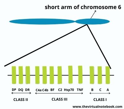
In humans, the HLA complex of genes is present on the short arm of chromosome 6 that contain several genes. These genes are critical to immune function. Further, MHC genes expressed co-dominantly in each individual. In other words, for a given major histocompatibility complex gene, each individual expresses alleles that will inherit from each of the two parents.
Types of Major Histocompatibility Complex
The HLA complex of genes is classified into three classes:
- Class I: HLA-A, HLA-B, and HLA-C.
- Class II: HLA-DR, HLA-DQ, and HLA-DP. All of these are present within the HLA-D region of the HLA complex.
- Class III: complement loci that encode for C2, C4, and factor B of the complement system and TNFs alpha and beta.
Class I and class II genes encode two groups of structurally distinct but homologous proteins. They also encode other nonpolymorphic genes whose products will involve in antigen presentation. Class I MHC molecules display peptides to CD8+ T cells whereas class II MHC molecules display peptides to CD4+ T cells. Class I and class II MHC molecules also differ in terms of which cells express them. Moreover, class I and class II MHC genes are the most polymorphic genes present in the genome.
Cellular distribution of MHC molecules
Class I MHC molecules are present in all nucleated cells with only two exceptions: neurons and striated muscle cells. They are particularly abundant on the surface of lymphocytes.
Class II MHC molecules are exclusively present in two groups of leukocytes. These are B lymphocytes and cells of the monocyte-macrophage family, which includes all antigen-presenting cells.
Major histocompatibility complex Structure
MHC molecules are membrane-bound glycoproteins that are closely related in both structure and function. These membrane glycoproteins function as highly specialized antigen-presenting molecules with grooves that form unusually stable complexes with peptide ligands.
Class I MHC molecules
These are glycoproteins present on the surface of virtually all nucleated cells. Class I proteins are encoded by the HLA-A, -B, and -C loci. Approximately, allelic genes encode 20 different proteins at the A locus, 40 at the B locus, and 8 at the C locus.
Class I molecules consist of two noncovalently linked polypeptide chains, an MHC-encoded 44- to 47-kD α chain (or heavy chain) and a non–MHC encoded 12-kD subunit called β2-microglobulin. All three molecules (class I chain, 2-microglobulin, and a peptide) are essential to the proper folding and expression of the MHC-peptide complex on the cell surface.
Basically, the heavy chain has a variable and constant region. The variable region is highly pleomorphic. The polymorphism of these molecules is important in the recognition of self and nonself. The heavy chain also has a constant region where the CD8 protein of the cytotoxic T cell binds. Most importantly, Class I proteins involved in graft rejection and cell-mediated cytolysis.
Class II MHC molecules
These are glycoproteins present on the surface of antigen-presenting cells, such as macrophages, B cells, dendritic cells of the spleen, and Langerhans’ cells of the skin. They are highly polymorphic glycoproteins comprises two noncovalently associated transmembrane of molecular weight of about 33,000 α chain and 29,000 Da β chain. The amino-terminal α1 and β1 segments of the class II chains interact to form the peptide-binding cleft, which is structurally similar to the cleft of class I molecules.
Unlike class I molecules, the genes encoding both chains of class II molecules are polymorphic and present in the MHC locus. The Class II MHC molecules contain external domains, a transmembrane segment, and a cytoplasmic anchor segment.
Class II proteins are primarily responsible for the graft-versus-host response and the mixed leukocyte response.
Class III MHC molecules
The class III MHC locus encodes complement proteins (C2, C4) of the classical pathways, properdin and factor B of the alternative pathway, and several cytokines.
General properties of MHC molecules
- Each MHC molecule consists of an extracellular peptide-binding cleft, or groove, followed by immunoglobulin (Ig)–like domains and transmembrane and cytoplasmic domains. Class I MHC molecules comprises two chains, one is MHC encoded and the other non-MHC encoded. On the other hand, class II MHC molecules have both MHC-encoded chains.
- The polymorphic amino acid residues of MHC molecules are present in and adjacent to the peptide-binding cleft. Because of amino acid variability in this region, different MHC molecules bind and display different peptides and are recognized specifically by the antigen receptors of different T cells.
- The nonpolymorphic Ig-like domains of MHC molecules contain binding sites for the T cell molecules CD4 and CD8. CD4 binds selectively to class II MHC molecules whereas CD8 binds to class I molecules.
Biological importance of MHC molecules
- MHC molecules bind peptide antigens and present them to T cells. Thus, these Major histocompatibility molecules are responsible for antigen recognition by the TCR (T-cell receptor). In this respect, the TCR is different from an antibody.
- Antibody molecules interact with antigen directly. Helper T cells recognize class II proteins. Helper cell activity depends on both the recognition of the antigen on antigen-presenting cells and the presence on these cells of “self” class II MHC proteins. This requirement to recognize antigen in association with a “self” MHC protein is called MHC restriction. MHC restriction in T cells occurs during their development in the thymus, specifically positive selection.
- MHC genes and proteins are also important in two other medical contexts. One is that many autoimmune diseases occur in people who carry certain MHC genes. Another is that the success of organ transplants depends on the compatibility of the MHC genes of the donor and recipient.
The function of Major histocompatibility complex
- MHC is the tissue-antigen that allows the immune system to bind to, recognize, and tolerate itself (autorecognition).
- Major histocompatibility complex also acts as the chaperone for intracellular peptides.
- MHC interacts with TCR and its co-receptors to optimize binding conditions for the TCR-antigen interaction and signal transduction effectiveness.
- They also involve in autoimmune reactions. Having some MHC molecules increases the risk of autoimmune diseases more than having others. HLA-B27 is an example. However, it is unclear how exactly it increases the risk of ankylosing spondylitis and other inflammatory diseases.
MHC and disease susceptibility
The MHC region is associated with more diseases than any other region of the human genome. In some cases, carrying specific HLA alleles may protect an individual against specific diseases. In other cases, carrying other HLA alleles may have unusual frequency of diseases generally classified as “autoimmune.”
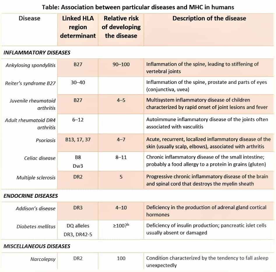
For example, individuals who do not express the class I HLA-Bw53 and the class II DQw5 alleles are at very high risk for the most severe and lethal forms of malaria. Nowadays, 40% of the natives of sub-Saharan Africa are positive for HLA-Bw53 and/or DQw5.
SOURCES AND EXTERNAL LINKS
Kuby immunology, 7th edition, chapter no. 8: Major histocompatibility complex and antigen representation.
Textbook of Microbiology and Immunology 2nd edition by Subhash Chandra Parija, chapter no. 16: structure and function of the immune system
Medical immunology, fifth edition revised and expanded, edited by Gabriel Virella, Major Histocompatibility complex
Review of medical microbiology and immunology by Warren Levinson, fourteenth edition, part VII; chapter no. 62
Lastly, Cellular and molecular immunology, 7th edition by Abul K. Abbas, MBBS chapter 6: Major histocompatibility complex molecules and antigen presentation to T lymphocytes
https://en.wikipedia.org/wiki/Major_histocompatibility_complex